The issue of post-printing editing is also a hot topic in 3D bioprinting, an increasingly popular tool in biomedical research and tissue engineering.
Growing tissues involves a highly complex environment of constantly changing signals to direct cell behaviour, requiring hydrogels with highly controllable mechanical properties. Editing 3D bioprinted hydrogels post-printing has posed a significant challenge, until a recent 2023 breakthrough by Falandt et al., published in Advanced Materials Technologies, unlocked new possibilities.
What is 3D bioprinting?

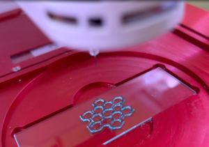
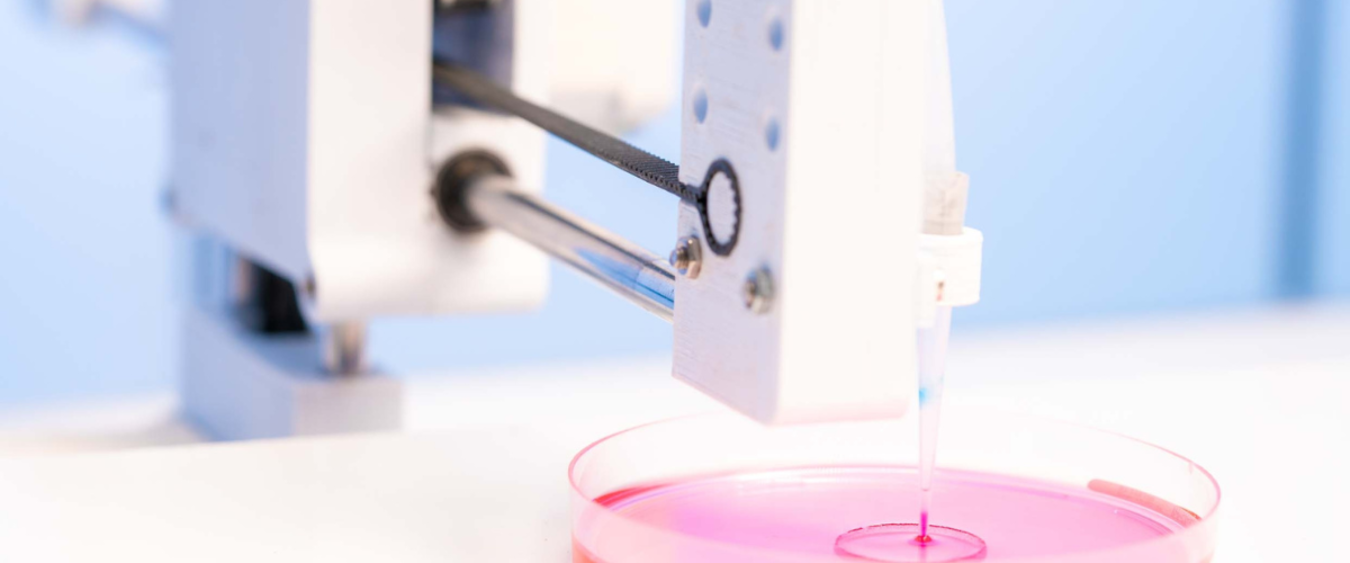
3D bioprinting is a similar concept to conventional 3D printing, in that a digital blueprint is converted into a physical object. However, bioprinting uses a special type of ink called bioink. These bioinks are a combination of biomaterials and living cells that can be printed, layer by layer, to create 3D cell models and tissue constructs. This technology gives precise control over where cells and biomolecules are positioned within the printed structure, meaning the extremely delicate shapes and structures of organs and tissues can be replicated to create fully functional tissue constructs.
3D bioprinting has many innovative medical applications, allowing the production of functional tissues for regenerative medicine, patient-specific models for drug development, and skin grafts and bone bandages for wound healing.
Post-printing editing – the challenge
In tissue engineering, 3D printed hydrogels must mimic the physicochemical composition of the natural extracellular matrix (ECM). However, the ECM plays a complex and active role, with region-dependent mechanical properties, and an array of biochemical signals that are released and presented in specific sequences to direct cell behaviour. Given this, the ideal 3D hydrogel would allow gradual presentation of different growth factors and morphogens into local cell environments, mimicking the natural changes occurring in development.
Currently, in conventional bioprinting, it’s very difficult to precisely edit the physicochemical properties after printing, preventing the programming of the time-dependent, on-demand cues that naturally developing cells would be exposed to in the body.
Post-printing editing – the solution
Falandt et al. have recently developed a new approach to help overcome the post-printing modification issue, publishing their findings in the journal Advanced Materials Technologies.
Using a technique called light-based volumetric printing, they can photograft thiolated compounds onto the hydrogel post-printing, with precise spatial patterning and custom geometries.

This light-based printing technique creates whole objects in a layerless fashion, allowing high-speed, high-resolution printing. Photografting can be done post-printing in a non-invasive and biocompatible manner, paving the way towards precise spatiotemporal modification of bioprinted constructs to better guide cell behaviour.
This approach is an important step towards creating biofabricated scaffolds that mimic the complex native biochemical environment of tissues and organs.
Unlocking new potential with transformative biomaterials
As a proof-of-concept, Falandt et al. photografted vascular endothelial growth factor into a bioprinted construct involving a gelatin norbornene (gelNOR) based biomaterial platform, produced from Rousselot’s X-Pure® gelatin.

Using Rousselot gelatin fulfilled a number of essential requirements:
- Assured biocompatibility
- Extremely low endotoxin content supporting translation for medical and pharmaceutical use
- Allows a wide range of chemical modifications
- Compatible with light-based 3D printing
- Tunable mechanical properties for varying levels of stiffness
- Supports photografting of any molecule with a free thiol group
Transformative biomaterials are a gateway to unlocking new possibilities in 3D bioprinting. The novel approach demonstrated by Falandt et al. shows that it is possible to control the spatial anchoring of biological cues through a hydrogel construct, taking us one step closer to the engineering of functional human tissues.
